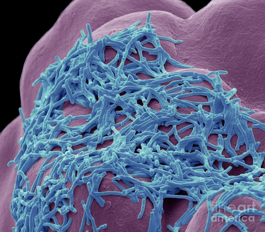
Mycobacterium smegmatis
A series of structome analyses, that is, quantitative and three-dimensional structural analysis of a whole cell at the electron microscopic level, have already been achieved individually in Exophiala dermatitidis, Saccharomyces cerevisiae, Mycobacterium tuberculosisMyojin spiral bacteria, and Escherichia coli. In these analyses, sample cells were processed through cryo-fixation and rapid freeze-substitution, resulting in the exquisite preservation of ultrastructures on the serial ultrathin sections examined by transmission mycobacterium smegmatis microscopy, mycobacterium smegmatis.
The genus Mycobacterium contains several slow-growing human pathogens, including Mycobacterium tuberculosis, Mycobacterium leprae, and Mycobacterium avium. Mycobacterium smegmatis is a nonpathogenic and fast growing species within this genus. In , a mutant of M. Classical bacterial models, such as Escherichia coli, were inadequate for mycobacteria research because they have low genetic conservation, different physiology, and lack the novel envelope structure that distinguishes the Mycobacterium genus. By contrast, M. Dissection and characterization of conserved genes, structures, and processes in genetically tractable M.
Mycobacterium smegmatis
Mycobacterium smegmatis is an acid-fast bacterial species in the phylum Actinomycetota and the genus Mycobacterium. It is 3. It was first reported in November by Lustgarten, who found a bacillus with the staining appearance of tubercle bacilli in syphilitic chancres. Subsequent to this, Alvarez and Tavel found organisms similar to that described by Lustgarten also in normal genital secretions smegma. This organism was later named M. Some species of the genus Mycobacterium have recently been renamed to Mycolicibacterium , so that M. Essentially, the bacteria form a single-layered sheet and are able to move slowly together without the use of any extracellular structures, like flagella or pili. For example, this sliding ability is correlated with the presence of glycopeptidolipids GLPs on the outermost part of the cell wall. GLPs are amphiphilic molecules that could potentially decrease surface interactions or create a conditioning film that allows movement. Although the exact role of GLPs in sliding is not known, without them M. Mycobacterium smegmatis is useful for the research analysis of other Mycobacteria species in laboratory experiments. The time and heavy infrastructure needed to work with pathogenic species prompted researchers to use M. Mycobacterium smegmatis shares the same peculiar cell wall structure of M. Bacterial secretion systems are specialized protein complexes and pathways that allow bacterial pathogens to secrete proteins across their cell membranes and, ultimately, to host cells. These effector proteins are important virulence factors, which allow the pathogen to survive inside of the host.
Cytoplasmic ribosome number was enumerated on each serial ultrathin section for seven cells. Rv Overexpression delays Mycobacterium smegmatis and Mycobacteria tuberculosis entry into mycobacterium smegmatis and increases pathogenicity of Mycobacterium smegmatis in mice.
.
A series of structome analyses, that is, quantitative and three-dimensional structural analysis of a whole cell at the electron microscopic level, have already been achieved individually in Exophiala dermatitidis, Saccharomyces cerevisiae, Mycobacterium tuberculosis , Myojin spiral bacteria, and Escherichia coli. In these analyses, sample cells were processed through cryo-fixation and rapid freeze-substitution, resulting in the exquisite preservation of ultrastructures on the serial ultrathin sections examined by transmission electron microscopy. In this paper, structome analysis of non pathogenic Mycolicibacterium smegmatis , basonym Mycobacterium smegmatis , was performed. Seven M. Cell profiles were measured, including cell length, diameter of cell and cytoplasm, surface area of outer membrane and plasma membrane, volume of whole cell, periplasm, and cytoplasm, and total ribosome number and density per 0. These data are based on direct measurement and enumeration of exquisitely preserved single cell structures in the transmission electron microscopy images, and are not based on the calculation or assumptions from biochemical or molecular biological indirect data. All measurements in M.
Mycobacterium smegmatis
The soil bacterium Mycobacterium smegmatis is able to scavenge the trace concentrations of H 2 present in the atmosphere, but the physiological function and importance of this activity is not understood. In this study, we explored the effect of deleting Hyd2 on cellular physiology by comparing the viability, energetics, transcriptomes, and metabolomes of wild-type vs. Hyd1 compensated for loss of Hyd2 when cells were grown in a high H 2 atmosphere. Analysis of cellular parameters showed that Hyd2 was not necessary to generate the membrane potential, maintain intracellular pH homeostasis, or sustain redox balance. We propose that atmospheric H 2 oxidation has two major roles in mycobacterial cells: to generate reductant during mixotrophic growth and to sustain the respiratory chain during dormancy. This is an open-access article distributed under the terms of the Creative Commons Attribution License , which permits unrestricted use, distribution, and reproduction in any medium, provided the original author and source are credited. Data Availability: The authors confirm that all data underlying the findings are fully available without restriction. All relevant data are within the paper and its Supporting Information files. The funders had no role in study design, data collection and analysis, decision to publish, or preparation of the manuscript. Competing interests: The authors have declared that no competing interests exist.
Gojol
These values are approximately twice of those of M. Amplification of Hsp 65 gene and usage of restriction endonuclease for identification of non-tuberculous rapid grower mycobacterium. Further to this analysis, it was revealed that M. It is 3. In Ziehl-Neelsen staining, M. The genus Mycobacterium contains several slow-growing human pathogens, including Mycobacterium tuberculosis, Mycobacterium leprae, and Mycobacterium avium. Read Edit View history. It depends on the RecA protein that catalyzes strand exchange and the ADN protein that acts as a presynaptic nuclease. In this paper, structome analysis of non pathogenic Mycolicibacterium smegmatis , basonym Mycobacterium smegmatis , was performed. Protein Sci. Smart specimen preparation for freeze substitution and serial ultrathin sectioning of yeast cells. The 3D reconstruction of Cell 3 with visualization of cell profile and cytoplasmic distribution of ribosomes. This suggests that if M.
Thank you for visiting nature. You are using a browser version with limited support for CSS. To obtain the best experience, we recommend you use a more up to date browser or turn off compatibility mode in Internet Explorer.
In addition, as shown in the following text, using exquisite TEM images obtained from serial ultrathin sections, three-dimensional reconstructions were performed. Brown-Elliott, B. Microscopy 66, — As shown in Table S1 , the cell diameters of M. Molecular mechanisms of intrinsic streptomycin resistance in Mycobacterium abscessus. Oren, A. On the contrary, ribosome density per 0. As discussed in previous literature, cell division of M. Then, cells with older growth poles elongate faster than cells with younger growth poles. Figure 3. Yang, K. Mycobacterium smegmatis is a nonpathogenic and fast growing species within this genus. Finally, our data strongly support the most recent establishment of the novel genus Mycolicibacterium , into which basonym Mycobacterium smegmatis has been classified. Essentially, the bacteria form a single-layered sheet and are able to move slowly together without the use of any extracellular structures, like flagella or pili. Srivastava, A.


The authoritative point of view, it is tempting
I congratulate, what words..., a brilliant idea
I am sorry, that I interfere, but I suggest to go another by.