Lipoma on pancreas
Pancreatic lipomas are thought to be very rare. Lipomas are usually easy to identify on imaging, particularly via computed tomography CT.
At the time the article was last revised Daniel J Bell had no financial relationships to ineligible companies to disclose. Pancreatic lipomas are uncommon mesenchymal tumors of the pancreas. Rarely symptomatic, they are most often detected incidentally on cross-sectional imaging for another purpose. If they do cause symptoms, it will typically be those related to regional mass effect from the mass. Pancreatic lipomas are composed of mature fat cells with thin internal fibrous septa. They differ from pancreatic lipomatosis in that they have well-defined margins covered by a thin collagen capsule.
Lipoma on pancreas
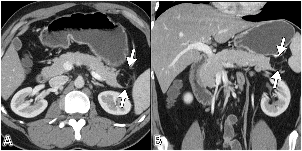
Federal government websites often end in. The site is secure. Pancreatic lipomas are rare. We present a case of incidentally discovered pancreatic lipoma in a year-old female suffering from metastatic ovarian carcinoma who was referred to radiology for follow-up imaging. Fat-containing tumours originating from the pancreas are very rare. Most lipomasshow characteristic features on imaging that allow their differentiation. In most cases, accurate diagnosis is attained without any histopathological confirmation. We present the imaging features of pancreatic lipoma on ultrasound, CT scan and MRI, the differential diagnosis and a brief review of the literature. On ultrasound imaging, lipomas are usually hyperechoic, although some lesions may demonstrate hypoechogenicity. Routine use of imaging and familiarity of the radiologists with this condition will increase the number of cases of pancreatic lipomas being diagnosed. The patient is a year-old female who was referred to the radiology department from the regional cancer center for imaging evaluation of a sonographically detected ovarian carcinoma. She was asymptomatic for the pancreatic lesion. She underwent CT imaging as a part of routine follow-up, which identified a pancreatic lipoma. Ultrasound and MRI were performed subsequently.
No ductal dilatation is apparent. She was treated symptomatically for biliary colic and was discharged well with a view for an elective laparoscopic cholecystectomy. Figure 3.
Regret for the inconvenience: we are taking measures to prevent fraudulent form submissions by extractors and page crawlers. Received: October 27, Published: November 27, Pancreatic lipoma and its differentiation from various fat containing lesions in the pancreas: an imaging guide. Int J Radiol Radiat Ther. DOI:
Federal government websites often end in. The site is secure. Recent studies have shown a significant increase in the utilization of computed tomography CT scans in the emergency department for a broad spectrum of conditions. This had a significant impact on the identification of patients with serious pathologies in a timely manner. However, the overutilization of computed tomography scans leads to increased identification of incidental findings. For example, pancreatic lesions are not uncommon findings that can be identified in imaging studies performed for other indications. Here, we report the case of a year-old male with a history of urinary stone disease who presented with right flank pain and dysuria. The urinalysis findings revealed numerous red blood cells and leukocytes.
Lipoma on pancreas
At the time the article was last revised Daniel J Bell had no financial relationships to ineligible companies to disclose. Pancreatic lipomas are uncommon mesenchymal tumors of the pancreas. Rarely symptomatic, they are most often detected incidentally on cross-sectional imaging for another purpose.
Upper credit humane society
They concluded that these show stable size, morphology and benign course, and only required short-term interval observation for proving their stability and differentiating them from early liposarcoma. Unable to process the form. CT appearance of incidental pancreatic lipomas: a case series. Retroperitoneal liposarcomas: the experience of a tertiary Asian center. Eur Radiol. Fat-containing tumours originating from the pancreas are very rare. Pathology also revealed chronic cholecystitis with cholesterol polyps. She followed regularly to the department of general surgery. Lipomatous tumors of the abdominal cavity: CT appearance and pathologic correlation. The final pathological examination confirmed a giant lipoma of the pancreas; the largest diameter was Suhas Aithal Sitharama: moc. URL of Article. Dei Tos AP.
Hence, localizing the tumor site can guide the healthcare provider to arrive at a probable diagnosis.
An abnormal 6. Loading Stack - 0 images remaining. It revealed a 6. This article is an open-access article which was selected by an in-house editor and fully peer-reviewed by external reviewers. Intrapancreatic lipoma and Morgagni hernia: a previously unrecognized association. Her medical history and family history were unremarkable. Case 1: lipoma in the head of the pancreas Case 1: lipoma in the head of the pancreas. The majority of them were asymptomatic at presentation and none of them required intervention, or showed interval growth or change on imaging appearance. In most of the reported cases, the lesions were located on the head of the pancreas, 3 — 5 , 11 , 12 , 14 — 16 as in our case. Fat-containing tumours originating from the pancreas are very rare. And a few fibroreticular septa could be seen within the lesion. MRI is very useful and shows almost total replacement of pancreatic parenchyma by fatty tissue.


0 thoughts on “Lipoma on pancreas”