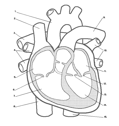
Unlabelled diagram of the heart
In this interactive, you can label parts of the human heart.
Diagram of the human heart, created by Wapcaplet in Sodipodi. Cropped by Yaddah to remove white space this cropping is not the same as Wapcaplet's original crop. Superior vena cava 2. Mitral valve 5. Aortic valve 6.
Unlabelled diagram of the heart
.
Inferior vena cava more wide. Go to full glossary Add 0 items to collection.
.
This file contains additional information such as Exif metadata which may have been added by the digital camera, scanner, or software program used to create or digitize it. If the file has been modified from its original state, some details such as the timestamp may not fully reflect those of the original file. The timestamp is only as accurate as the clock in the camera, and it may be completely wrong. From Wikimedia Commons, the free media repository. File information. Structured data. Captions Captions English Add a one-line explanation of what this file represents.
Unlabelled diagram of the heart
The major organs of the respiratory system function primarily to provide oxygen to body tissues for cellular respiration, remove the waste product carbon dioxide, and help to maintain acid-base balance. Portions of the respiratory system are also used for non-vital functions, such as sensing odors, speech production, and for straining, such as during childbirth or coughing Figure Functionally, the respiratory system can be divided into a conducting zone and a respiratory zone. The conducting zone of the respiratory system includes the organs and structures not directly involved in gas exchange. The gas exchange occurs in the respiratory zone. The major functions of the conducting zone are to provide a route for incoming and outgoing air, remove debris and pathogens from the incoming air, and warm and humidify the incoming air. Several structures within the conducting zone perform other functions as well. The epithelium of the nasal passages, for example, is essential to sensing odors, and the bronchial epithelium that lines the lungs can metabolize some airborne carcinogens. The major entrance and exit for the respiratory system is through the nose. When discussing the nose, it is helpful to divide it into two major sections: the external nose, and the nasal cavity or internal nose.
Reddit post karma vs comment karma
Mitral valve 5. Derivative works of this file: Fontan procedure. Yes No. File Talk. This survey will open in a new tab and you can fill it out after your visit to the site. Selecting or hovering over a box will highlight each area in the diagram. Description Diagram of the human heart cropped. I, the copyright holder of this work, hereby publish it under the following licenses:. The following other wikis use this file: Usage on ar. Cropped by Yaddah to remove white space this cropping is not the same as Wapcaplet's original crop. The source code of this SVG is invalid due to an error. Add papillary muscles and chordae tendinae.
The right side pumps deoxygenated close deoxygenated Blood that is low in oxygen as cells have used it and high in carbon dioxide as cells have produced it. The left side pumps oxygenated close oxygenated Blood that is high in oxygen and low in carbon dioxide. This unidirectional flow of blood through the heart shows that mammals have a double circulatory system.
Drag and drop the text labels onto the boxes next to the heart diagram. Aorta Labelling the heart The heart is a muscular organ that pumps blood through the blood vessels of the circulatory system. Creative Commons Attribution-ShareAlike 3. Appears in. Parts of the heart Labels Description vena cava Carries deoxygenated blood from the body to the heart semilunar valve Flaps that prevent backflow of blood left atrium Receives oxygenated blood from the lungs left ventricle Region of the heart that pumps oxygenated blood to the body pulmonary artery Carries deoxygenated blood to the lungs right ventricle Region of the heart that pumps deoxygenated blood to the lungs pulmonary vein Carries oxygenated blood from the lungs right atrium Segment of the heart that receives deoxygenated blood aorta The main artery carrying oxygenated blood to all parts of the body. Use Reset All to practise again from the start. Download 0 items. English: Diagram of the human heart 1. The source code of this SVG is invalid due to an error. Cropped by Yaddah to remove white space this cropping is not the same as Wapcaplet's original crop.


I am sorry, it not absolutely approaches me. Who else, what can prompt?
It is interesting. You will not prompt to me, where to me to learn more about it?