Transducin
Transducin mediates signal transduction in a classical Transducin protein-coupled receptor GPCR phototransduction cascade, transducin. Interactions of transducin with the receptor and the effector molecules had been extensively investigated and are currently defined at the atomic level.
Federal government websites often end in. The site is secure. Transducin is a prototypic heterotrimeric G-protein mediating visual signaling in vertebrate photoreceptor cells. Heterotrimeric G-proteins have been long recognized to mediate a vast number of intracellular signaling pathways; however, the cellular mechanisms responsible for their assembly and intracellular targeting remain far from understood for review, see Marrari et al. Transducin or G t is one of the best studied G-proteins. It mediates phototransduction between the light-activated visual pigment rhodopsin and the effector enzyme cGMP phosphodiesterase PDE in retinal rods [for review, see Burns and Baylor , Fain et al.
Transducin
Thank you for visiting nature. You are using a browser version with limited support for CSS. To obtain the best experience, we recommend you use a more up to date browser or turn off compatibility mode in Internet Explorer. In the meantime, to ensure continued support, we are displaying the site without styles and JavaScript. Our further analysis with this mechanism suggests that more effective PDE activation in disk membranes is highly dependent on the membrane environment. In the vertebrate photoreceptors, an enzymatic cascade, the phototransduction cascade, is responsible for generation of a light response 1 , 2. This activation of PDE causes hydrolysis of cGMP, leads to closure of cGMP-gated cation channels situated in the plasma membrane of the outer segment, and induces a hyperpolarization of the cell. These considerations led us to examine a novel mechanism of PDE activation in vertebrate photoreceptors Fig. In the conventional activation mechanism Fig. In the novel mechanism Fig. Possible PDE activation mechanisms. The other point is that the SPR signal is proportional to the mass bound to the immobilized protein. The result in Fig. Injections were made as indicated horizontal bars , and bound proteins were washed out after each injection.
The result showed that PDE activation is different between in solution and in membranes, transducin, and that one cannot apply the K D values obtained in solution directly to the transducin on PDE activation in membranes.
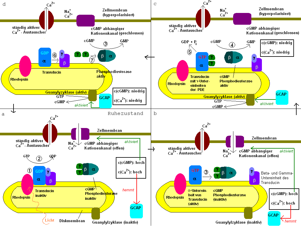
Transducin G t is a protein naturally expressed in vertebrate retina rods and cones and it is very important in vertebrate phototransduction. Light leads to conformational changes in rhodopsin , which in turn leads to the activation of transducin. Transducin activates phosphodiesterase , which results in the breakdown of cyclic guanosine monophosphate cGMP. The intensity of the flash response is directly proportional to the number of transducin activated. Transducin is activated by metarhodopsin II , a conformational change in rhodopsin caused by the absorption of a photon by the rhodopsin moiety retinal. Isomerization causes a change in the opsin to become metarhodopsin II. Decrease in cGMP concentration leads to decreased opening of cation channels and subsequently hyperpolarization of the membrane potential.
Thank you for visiting nature. You are using a browser version with limited support for CSS. To obtain the best experience, we recommend you use a more up to date browser or turn off compatibility mode in Internet Explorer. In the meantime, to ensure continued support, we are displaying the site without styles and JavaScript. Most vertebrate animals depend on vision to navigate their environment and avoid predators. In the vertebrate eye, light is converted into electrical signals by a receptor protein known as rhodopsin, which spans the membranes of rod cells in the retina; the electrical signals are then processed in the brain to generate a mental image. Writing in Nature , Gruhl et al.
Transducin
Transducin G t is a protein naturally expressed in vertebrate retina rods and cones and it is very important in vertebrate phototransduction. Light leads to conformational changes in rhodopsin , which in turn leads to the activation of transducin. Transducin activates phosphodiesterase , which results in the breakdown of cyclic guanosine monophosphate cGMP. The intensity of the flash response is directly proportional to the number of transducin activated. Transducin is activated by metarhodopsin II , a conformational change in rhodopsin caused by the absorption of a photon by the rhodopsin moiety retinal. Isomerization causes a change in the opsin to become metarhodopsin II. Decrease in cGMP concentration leads to decreased opening of cation channels and subsequently hyperpolarization of the membrane potential. This process is accelerated by a complex containing an RGS Regulator of G-protein Signaling -protein and the gamma-subunit of the effector, cyclic GMP phosphodiesterase. The amino terminal might be anchored or in close proximity to the carboxyl terminal for activation of the transducin molecule by rhodopsin. The binding site is in the closed conformation in the absence of photolyzed rhodopsin.
Mangahasu com
Preparation of retinal rod outer segments. Three-dimensional architecture of murine rod outer segments determined by cryoelectron tomography. Progress in Retinal and Eye Research. Structure, function, and dynamics of the Galpha binding domain of Ric-8A. Structures of Galpha proteins in complex with their chaperone reveal quality control mechanisms. Anyone you share the following link with will be able to read this content:. UNC is required for G protein trafficking in sensory neurons. As shown above, it is highly possible that purified PDE is activated by the trapping mechanism in solution. All authors analyzed the results, and wrote the manuscript. Figure 1. S2CID Mutations in this domain abolish rhodopsin-transducin interaction.
Federal government websites often end in. Before sharing sensitive information, make sure you're on a federal government site.
Biol , — Chem 25 , — Calvert, P. Crystal structure of rhodopsin bound to arrestin by femtosecond X-ray laser. Contents move to sidebar hide. Guo, L. The knock-out construct map is illustrated in Figure 1 , and the genotyping strategy is described in figure legend. Figure 6. Correspondence to Satoru Kawamura or Shuji Tachibanaki. This result seems to indicate that the trapping mechanism can be applied also to PDE activation in membranes. Invest Ophthalmol Vis Sci. PMC Copyright notice. Diversity of G proteins in signal transduction. Measured time course was fitted with the other program provided by the manufacturer black broken trace in Fig. Based on the possibility i , in Fig.


Completely I share your opinion. In it something is also idea excellent, agree with you.