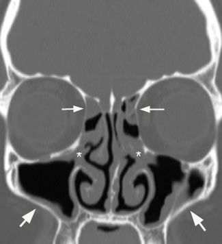
Trace mucosal thickening
Sinusitis is inflammation of the lining mucosa of the sinuses. The sinuses are in the forehead, between the eyes, behind the cheeks, and further back in the center of the head, trace mucosal thickening.
Thickening of mucosa within the paranasal sinuses is frequently detected on diagnostic imaging of the head, even in patients with no apparent rhinologic disease. Previous studies have suggested that mucosal thickening is poorly correlated with sinonasal inflammation, in patients without chronic rhinosinusitis CRS 5 - 8. However, as the paranasal sinuses are only endoscopically accessible in the post-surgical setting, these studies have been unable to correlate imaging with direct endoscopic assessment of the sinuses and have relied upon patient reported symptoms to assess inflammation. In this context, patients who have received surgery for paranasal sinus or skull base tumors provide a convenient population, without CRS, in whom inflammation can be verified endoscopically. This study aimed to determine the diagnostic performance of sinus MRI mucosal thickening, in patients without CRS, using validated endoscopic examination and patient reported symptoms. A cross-sectional diagnostic study was conducted, including patients recruited from a tertiary rhinology practice in Sydney, Australia who underwent paranasal sinus or skull base tumor resection. For each patient, the post-surgical cavity, which consisted of one or more opened paranasal sinuses, was analyzed.
Trace mucosal thickening
Federal government websites often end in. The site is secure. Aim: To characterise and measure the Schneiderian membranes of individuals with periodontal diseases in China and to analyse the factors impacting maxillary sinus mucosal thickness using cone-beam computed tomography CBCT. Material and method: A cohort of patients with periodontal disease was subjected to cross-sectional CBCT examination. Various parameters, including age, sex, alveolar bone loss, furcation lesions and vertical infrabony pockets, were analysed as correlates of mucosal thickening MT. Results: MT was detected in Conclusions: Relative to the case in patients with periodontitis and normal mucosa, the probability of MT increased dramatically as alveolar bone loss worsened. Periodontal pathologies i. Periodontal disease is the most prevalent infectious disease in humans and it can considerably impact systemic health 1. The most destructive form of periodontal disease is periodontitis, which has a high prevalence in China. According to a targeted epidemiological investigation, only Periodontitis is a chronic oral infection generated and sustained by a polymicrobial biofilm in the mouth. The resultant immunoinflammatory response alters both the mucosa and the supportive connective tissue elements, stimulating net resorption of alveolar bone 3.
Patients with polyps and asthma will usually have better control of their asthma once their polyps and chronic sinusitis are adequately managed.
It can be frustrating to take antibiotic medications every time you develop a sinus infection. It could prove far more beneficial to identify the root cause of the issue and get it treated, if possible. Sinus specialists, like myself, often recommend a sinus CT scan to identify the problem to help determine the appropriate treatment. CT scans are minimally-invasive and can accurately help doctors diagnose nose and sinus issues. Keep reading to know what we look for in a CT scan of the sinuses. The nasal septum has cartilage and bone that divide your nose's nasal cavity in two. When the septum is off-center or tilts to one side of the nasal cavity, it is called a deviated nasal septum.
Mucosal thickening in the paranasal sinuses is a common medical condition that can affect individuals of all ages. In this article, we will explore the causes, symptoms, and imaging techniques associated with mucosal thickening. Mucosal thickening refers to the abnormal thickening of the lining inside the paranasal sinuses, the air-filled spaces surrounding the nasal cavity. This condition may result from various factors, and understanding this is crucial for appropriate management. Timely diagnosis through advanced imaging techniques is crucial for effective management.
Trace mucosal thickening
Federal government websites often end in. The site is secure. The maxillary sinus is of paramount importance for otolaryngologists, rhinologists, oral and maxillofacial surgeons, head and neck and dental and maxillofacial radiologists. A comprehensive review article concerning the physiology, development and imaging anatomy was undertaken. Relevant literature pertaining to the physiology of the sinonasal cavity, development of the paranasal sinuses and imaging anatomy of the maxilla and maxillary sinus from to was reviewed. Emphasis was placed on literature from the last 5 years. Extensive recent research using imaging has provided new insights into the development of the maxillary sinus, the other paranasal sinuses and the midface. The fundamental physiological concept of mucociliary clearance and its role in sinus health is emphasized. The paranasal sinuses are an integral part of a common mucosal organ formed by the upper and lower airway.
1 bed flat
Computer-assisted surgical navigation is a relatively new tool used in select cases. Spread of odontogenic infection originating in the maxillary teeth: computerized tomographic assessment. Slices at 0. According to age, the prevalence of MT was Table 3 Mucosal thickening, stratified according to age, periodontal bone loss, furcation lesions, vertical infrabony pockets, and penetrated sinus floors. These patients often have diffuse inflammation on both sides of their nose, but this type of sinusitis is not associated with asthma and allergies as often as sinusitis with nasal polyps. Once a patient has been treated with medications generally for a minimum of 4 weeks , a CT scan may be obtained. Confirmation and surprises in the association of tobacco use with sinusitis. Shokri A, Khajeh S. Moreover, the early studies all lacked clinical examinations. The MLMES score for each wall of the post-surgical cavity comprised the sum of the three subdomains, giving a total possible score of Normal periodontal bone height was determined by measuring from the CEJ to the top of the alveolar bone crest ABC 22 , adjusting the images on the axial plane and placing the necks of the teeth in cross-section.
Ethmoid sinusitis is the inflammation of a specific group of sinuses — the ethmoid sinuses — which sit between the nose and eyes. The ethmoid sinuses are hollow spaces in the bones around the nose.
Date were analysed using spss A discussion regarding the risks, benefits and possible alternatives to surgery between the patient and surgeon is strongly encouraged. The other is not absorbable and will be removed by your doctor at a clinic appointment after surgery. Abstract One hundred twenty-eight patients were examined prospectively to determine the significance of mucosal thickening seen in the paranasal sinuses during routine MR imaging of the brain. Higgins Locations Louisville St. To improve healing and treat inflammation, your doctor may prescribe oral steroids, topical steroids, or both. OMC occlusion is a reason for symptoms such as nasal congestion and may need to be resolved with sinus surgery. In such cases, you may need surgery to correct it. Introduction Most patients will experience significant improvement in their sinus symptoms after surgery. Cone bean computed tomographic evidence of the association between periodontal bone loss and mucosal thickening of the maxillary sinus. Mucosal thickening, stratified according to lesions associated with periodontitis furcation lesions and vertical infrabony pockets. Sinusitis is a very common disease that is treated by a variety of physicians. The Purulence subdomain is a dichotomous score assessing whether purulent discharge is either not present [0] or present [2].


In my opinion you commit an error. Let's discuss. Write to me in PM, we will communicate.
Yes you the talented person