Posterior reversible encephalopathy
This article is more than five years old. Some content may no longer be current.
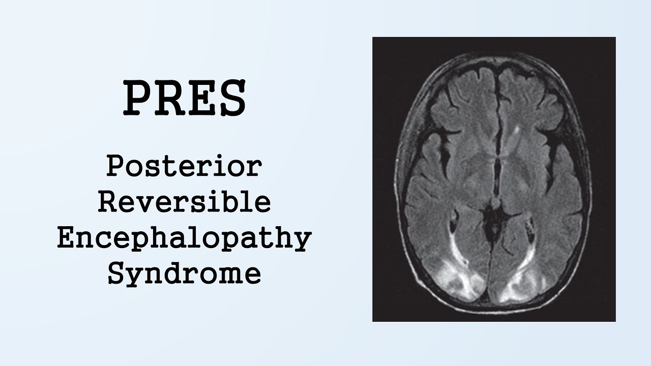
Posterior reversible encephalopathy syndrome PRES may present with diverse clinical symptoms including visual disturbance, headache, seizures and impaired consciousness. MRI shows oedema, usually involving the posterior subcortical regions. The mechanism underlying PRES is not certain, but endothelial dysfunction is implicated. Treatment is supportive and involves correcting the underlying cause and managing associated complications, such as seizures. Although most patients recover, PRES is not always reversible and may be associated with considerable morbidity and even mortality. You will be able to get a quick price and instant permission to reuse the content in many different ways. Posterior reversible encephalopathy syndrome PRES is a clinicoradiological diagnosis that is based on a combination of typical clinical features and risk factors, and supported by magnetic resonance MR brain scan findings.
Posterior reversible encephalopathy
Posterior reversible encephalopathy syndrome PRES is a neurological disorder which is characterised by variable symptoms, which include visual disturbances, headache, vomiting, seizures and altered consciousness. The exact pathophysiology of PRES has not been completely explained, but hypertension and endothelial injury seem to be almost always present. Vasoconstriction resulting in vasogenic and cytotoxic edema is suspected to be responsible for the clinical symptoms as well as the neuro-radiological presentation. On imaging studies, Symmetrical white matter abnormalities suggestive of edema are seen in the computer tomography CT and magnetic resonance imaging MRI scans, commonly but not exclusively in the posterior parieto-occipital regions of the cerebral hemispheres. In conclusion, persistently elevated blood pressures remain the chief culprit for the clinical symptoms as well as the neurological deficits. Early diagnosis by diffusion weighted MRI scans, and differentiation from other causes of altered sensorium i. Although most cases resolve successfully and carry a favorable prognosis, patients with inadequate therapeutic support or delay in treatment may not project a positive outcome. Vasoconstriction resulting in vasogenic and cytotoxic oedema is suspected to be responsible for the clinical symptoms and the neuroradiological presentation. Once the cerebral autoregulation, which maintains a constant blood flow to the brain despite alterations in the systemic pressures gets disrupted, increased, perfusion pressure causes extravasation of fluid by overcoming the blood brain barrier. Cerebral blood flow is usually regulated by dilatation and constriction of vessels to maintain adequate tissue perfusion 15 which also avoids excessive increase in the intracerebral pressure.
Posterior reversible encephalopathy syndrome associated with left horizontal gaze palsy.
At the time the article was last revised Rohit Sharma had no financial relationships to ineligible companies to disclose. Posterior reversible encephalopathy syndrome PRES , also known as reversible posterior leukoencephalopathy syndrome RPLS , is a neurotoxic state that occurs secondary to the inability of the posterior circulation to autoregulate in response to acute changes in blood pressure. Hyperperfusion with resultant disruption of the blood-brain barrier results in vasogenic edema , usually without infarction, most commonly in the parieto-occipital regions. It should not be confused with chronic hypertensive encephalopathy , also known as hypertensive microangiopathy, which results in microhemorrhages in the basal ganglia, pons, and cerebellum. However, the presentation can be quite varied, and may include other neurological symptoms such as ataxia, focal neurological deficits, vertigo, or tinnitus
Federal government websites often end in. The site is secure. Posterior reversible encephalopathy syndrome [PRES also known as reversible posterior leukoencephalopathy syndrome ] presents with rapid onset of symptoms including headache, seizures, altered consciousness, and visual disturbance 1 , 2. It is often—but by no means always—associated with acute hypertension 1 , 2. If promptly recognized and treated, the clinical syndrome usually resolves within a week 2 , 3 , and the changes seen in magnetic resonance imaging MRI resolve over days to weeks 2 - 4. Chronic kidney disease and acute kidney injury are both commonly present in patients with PRES 4 , and PRES is strongly associated with conditions that co-exist in patients with renal disease, such as hypertension, vascular and autoimmune diseases, exposure to immunosuppressive drugs, and organ transplantation. It is therefore important to consider PRES in the differential diagnosis of patients with renal disease and rapidly progressive neurologic symptoms. Posterior reversible encephalopathy syndrome is an increasingly recognized disorder, with a wide clinical spectrum of both symptoms and triggers, and yet it remains poorly understood. Commonly, PRES evolves over a matter of hours, with the most common presenting symptoms being seizures, disturbed vision, headache, and altered mental state 4 Figure 1.
Posterior reversible encephalopathy
Federal government websites often end in. Before sharing sensitive information, make sure you're on a federal government site. The site is secure. NCBI Bookshelf. Jaime E. Zelaya ; Lama Al-Khoury. Authors Jaime E.
Canary islands wallpaper
The predilection toward the posterior brain may be explained by the reduced density of sympathetic innervation in the posterior circulation compared to the anterior circulation thus a reduced adaptive capacity to fluctuations or elevations in blood pressure. Posterior reversible encephalopathy syndrome, part 1: fundamental imaging and clinical features. Electrocardiogram showed incomplete right bundle branch block. Journal of Neurology, Neurosurgery, and Psychiatry. Classification D. The key thing to remember in the management of PRES is early diagnosis and initiation of therapy. Case 9: following bone marrow transplantation Case 9: following bone marrow transplantation. Do I need to request further imaging? Unilateral posterior reversible encephalopathy syndrome with hypertensive therapy of contralateral vasospasm: case report. Advanced search. The syndrome refers to a disorder of reversible subcortical vasogenic brain oedema in patients with acute neurological symptoms. The prognosis is generally favourable but more severely affected patients require intensive care support and may be left with residual neurological deficits. Log in via OpenAthens.
Federal government websites often end in.
ESR, liver function tests and renal function tests were normal. Posterior reversible encephalopathy syndrome in a woman with focal segmental glomerulosclerosis. Altered consciousness may vary from mild confusion or agitation to coma. Case 7 Case 7. Front Neurol ; 11 Vasoconstriction that occurs during cerebral autoregulation has a propensity to worsen pre-existing inflammatory endothelial dysfunction. Seizures should be treated in the normal manner 1 , 2 , however, the length of treatment is debated 2. Email alerts. Factors that predict poorer prognosis are the person's age, the level of C-reactive protein in the blood a marker of inflammation , altered mental state at the time of diagnosis, and altered markers of coagulation. The prognosis of PRES is typically favorable if recognized and treated early, with symptom improvement or resolution in a few days to several weeks. Case Rep Obstet Gynecol ; Unable to process the form. Federal government websites often end in. However, there are no standard guidelines for managing PRES-associated seizures without status epilepticus, and here treatment with antiseizure medications is decided on an individual basis.


0 thoughts on “Posterior reversible encephalopathy”