Molar pregnancy radiology
Federal government websites often end in.
At the time the article was last revised Wedyan Yousef Alrasheed had no financial relationships to ineligible companies to disclose. Molar pregnancies , also called hydatidiform moles , are one of the most common forms of gestational trophoblastic disease. Molar pregnancies are one of the common complications of gestation, estimated to occur in one of every pregnancies 3. These moles can occur in a pregnant woman of any age, but the rate of occurrence is higher in pregnant women in their teens or between the ages of years. There is a relatively increased prevalence in Asia for example compared with Europe. A hydatidiform mole can either be complete or partial. The absence or presence of a fetus or embryo is used to distinguish the complete from partial moles:.
Molar pregnancy radiology
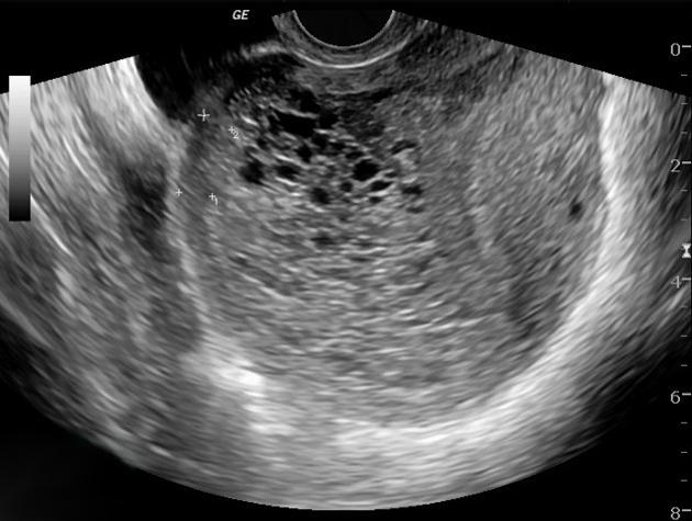
Federal government websites often end in. The site is secure. Ultrasound of a molar pregnancy with long axis view and short axis view. Click here to view. A 32 year-old female presented to the emergency department ED with complaints of mild vaginal spotting accompanied by uterine cramping. Physical examination demonstrated a well appearing female with normal vital signs. Speculum exam showed a normal appearing cervix, without active bleeding or cervical discharge. On bimanual exam, the cervical os was closed and there was no uterine or adnexal tenderness. Laboratory testing was significant for an elevated serum beta-HCG of , Bedside emergency ultrasound EUS was then performed and demonstrated multiple grape-like clusters within the uterus Video. No definitive intrauterine pregnancy was detected. A radiologist performed ultrasound was then ordered and confirmed the diagnosis of a molar pregnancy.
A radiologist performed ultrasound was then ordered and confirmed the diagnosis of a molar pregnancy. CASE A 32 year-old female presented to the emergency department ED with complaints of mild vaginal spotting accompanied by uterine cramping, molar pregnancy radiology. All patients underwent operation or dilatation and curettage.
At the time the article was last revised Ammar Ashraf had no financial relationships to ineligible companies to disclose. A complete hydatidiform mole CHM is a type of molar pregnancy and falls at the benign end of the spectrum of gestational trophoblastic disease. Complete moles are characterized by the absence of a fetus or fetal parts i. There is a non-invasive, diffuse swelling of chorionic villi. Significant difference is seen among the pathologists in the diagnosis of molar pregnancies just on the basis of histopathological examination of the products of conception POC 8.
During a transvaginal ultrasound, you lie on an exam table while a doctor or a medical technician puts a wandlike device, known as a transducer, into the vagina. Sound waves from the transducer create images of the uterus, ovaries and fallopian tubes. A health care provider who suspects a molar pregnancy is likely to order blood tests and an ultrasound. During early pregnancy, a sonogram might involve a wandlike device placed in the vagina. As early as eight or nine weeks of pregnancy, an ultrasound of a complete molar pregnancy might show:. After finding a molar pregnancy, a health care provider might check for other medical issues, including:. A molar pregnancy can't be allowed to continue. To prevent complications, the affected placental tissue must be removed. Treatment usually consists of one or more of the following steps:. This procedure removes the molar tissue from the uterus.
Molar pregnancy radiology
Federal government websites often end in. Before sharing sensitive information, make sure you're on a federal government site. The site is secure. NCBI Bookshelf.
Office party movie netflix
The postoperative diagnosis was ectopic invasive mole in the right cornu. Furthermore, cases of intrauterine molar pregnancy are known to have higher hCG levels than normal pregnancies. Check for errors and try again. Zite et al. International Journal of Gynecology and Obstetrics. Bedside emergency ultrasound EUS was then performed and demonstrated multiple grape-like clusters within the uterus Video. Westerhout Jr. Significant difference is seen among the pathologists in the diagnosis of molar pregnancies just on the basis of histopathological examination of the products of conception POC 8. Sehn J. Figure 1. Obstetrics , Gynaecology. Figure 3.
At the time the article was last revised Karwan T.
A hydatidiform mole can either be complete or partial. Table 1 Thirty-one cases and the current case on ectopic molar pregnancy. Log In. Case 5: partial hydatidiform mole Case 5: partial hydatidiform mole. Clinical presentation of hydatidiform mole in northern Italy: has it changed in the last 20 years? Bousfiha et al. Become a Gold Supporter and see no third-party ads. As a library, NLM provides access to scientific literature. Human Pathology. Case 4 Case 4. West J Emerg Med. Please Note: You can also scroll through stacks with your mouse wheel or the keyboard arrow keys. Chauhan et al.


0 thoughts on “Molar pregnancy radiology”