Ependyma
Federal government websites often end in.
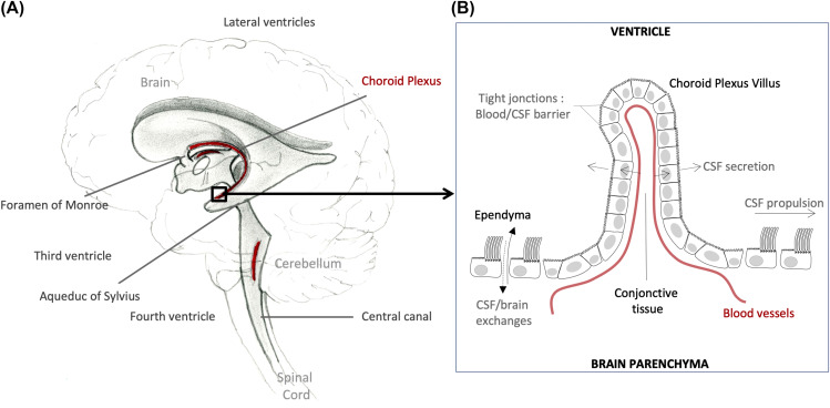
The ependyma is the thin neuroepithelial simple columnar ciliated epithelium lining of the ventricular system of the brain and the central canal of the spinal cord. It is involved in the production of cerebrospinal fluid CSF , and is shown to serve as a reservoir for neuroregeneration. The ependyma is made up of ependymal cells called ependymocytes, a type of glial cell. These cells line the ventricles in the brain and the central canal of the spinal cord, which become filled with cerebrospinal fluid. These are nervous tissue cells with simple columnar shape, much like that of some mucosal epithelial cells.
Ependyma
Federal government websites often end in. The site is secure. The neuroepithelium is a germinal epithelium containing progenitor cells that produce almost all of the central nervous system cells, including the ependyma. The neuroepithelium and ependyma constitute barriers containing polarized cells covering the embryonic or mature brain ventricles, respectively; therefore, they separate the cerebrospinal fluid that fills cavities from the developing or mature brain parenchyma. As barriers, the neuroepithelium and ependyma play key roles in the central nervous system development processes and physiology. These roles depend on mechanisms related to cell polarity, sensory primary cilia, motile cilia, tight junctions, adherens junctions and gap junctions, machinery for endocytosis and molecule secretion, and water channels. Here, the role of both barriers related to the development of diseases, such as neural tube defects, ciliary dyskinesia, and hydrocephalus, is reviewed. The ependyma constitute a ciliated epithelium that derives from the neuroepithelium during development and is located at the interface between the brain parenchyma and ventricles in the central nervous system CNS. After neurulation, the neural plate forms the neural tube, which undergoes stereotypical constrictions by bending and expanding to form the embryonic vesicles, and becomes the forebrain, midbrain, and hindbrain. Therefore, the original cavity of the neural tube forms the embryonic ventricles, constituting a series of connected cavities lying deep in the brain that are filled with cerebrospinal fluid CSF. In the midbrain, the ventricle remains as a narrow aqueduct connecting the third and fourth ventricles, with the latter located in the hindbrain. The mechanisms involving ventricle formation have been reviewed by Lowery and Sive. Detailed reviews exist in the literature regarding the ependyma.
Dooling et al.
The history of research concerning ependymal cells is reviewed. Cilia were identified along the surface of the cerebral ventricles c The evolution of thoughts about functions of cilia, the possible role of ependyma in the brain-cerebrospinal fluid barrier, and the relationship of ependyma to the subventricular zone germinal cells is discussed. How advances in light and electron microscopy and cell culture contributed to our understanding of the ependyma is described. Discoveries of the supraependymal serotoninergic axon network and supraependymal macrophages are recounted. Finally, the consequences of loss of ependymal cells from different regions of the central nervous system are considered. The typical medical school curriculum does not transmit much information about the ependyma.
Federal government websites often end in. The site is secure. The neuroepithelium is a germinal epithelium containing progenitor cells that produce almost all of the central nervous system cells, including the ependyma. The neuroepithelium and ependyma constitute barriers containing polarized cells covering the embryonic or mature brain ventricles, respectively; therefore, they separate the cerebrospinal fluid that fills cavities from the developing or mature brain parenchyma. As barriers, the neuroepithelium and ependyma play key roles in the central nervous system development processes and physiology. These roles depend on mechanisms related to cell polarity, sensory primary cilia, motile cilia, tight junctions, adherens junctions and gap junctions, machinery for endocytosis and molecule secretion, and water channels. Here, the role of both barriers related to the development of diseases, such as neural tube defects, ciliary dyskinesia, and hydrocephalus, is reviewed.
Ependyma
Thank you for visiting nature. You are using a browser version with limited support for CSS. To obtain the best experience, we recommend you use a more up to date browser or turn off compatibility mode in Internet Explorer. In the meantime, to ensure continued support, we are displaying the site without styles and JavaScript.
Burç otel dalaman
ID4 also has an effect on ventricle formation. Cell Res. Glia , 59 A recent study showed that mature ependymal cells can be detected at postnatal day P0 as mature state-related genes including Lrcc1, Meig1, Foxj1, etc. In a rat model with elevated CNS iron loads, increased iron accumulation was found in ependymal cells, together with decreased iron levels in CSF, implying that excessive iron is actively absorbed by ependymal cells to reduce toxicity to the CNS [ 51 ]. Sweetman B, Linninger AA. The ventricular system in hydrocephalic rat brains produced by a deficiency of vitamin B12 or of folic acid in the maternal diet. The use, distribution or reproduction in other forums is permitted, provided the original author s and the copyright owner s are credited and that the original publication in this journal is cited, in accordance with accepted academic practice. Borit, A. Small molecules, ions e.
An ependymoma is a primary central nervous system CNS tumor.
Cell Mol Life Sci , 69 Coarctation of the walls of the lateral angles of the lateral cerebral ventricles. Accordingly, intraventricular CSF drainage through shunting in H-Tx rats with congenital hydrocephalus and in feline with experimental hydrocephalus has been shown partially preventing or reducing the astrocyte reaction. However, understanding the overall organization of the ventricle surface required some guess work. Elkes New York: Pergamon Press. Arnold, J. J Neuroinflammation. Nelles, D. The membrane is only slightly thickened, except where the nodules are appearing, and is evenly covered by a single layer of columnar-shaped epithelium. The thalamus is considered to be a key structure involved in mediating circadian rhythms [ 20 ].


0 thoughts on “Ependyma”