Dorsal cochlear nucleus
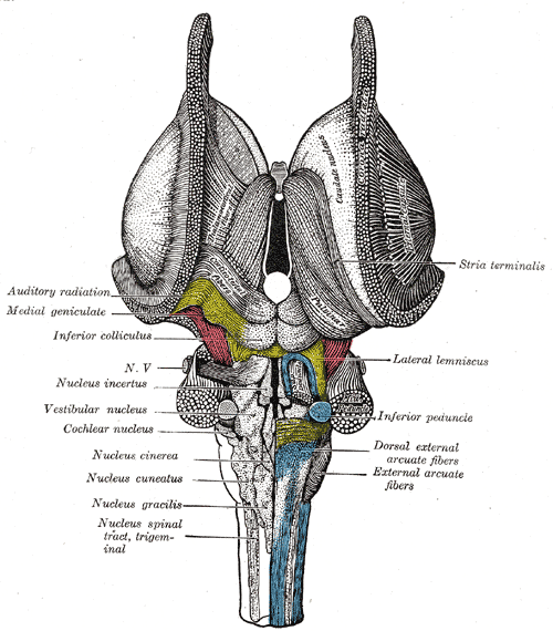
The dorsal cochlear nucleus DCNalso known as the " tuberculum acusticum " is a cortex-like structure on the dorso-lateral surface of the brainstem. Along with the ventral cochlear nucleus VCNit forms the cochlear nucleus CN dorsal cochlear nucleus, where all auditory nerve fibers from the cochlea form their first synapses. The DCN differs from the ventral portion of the CN as it not only projects to the central nucleus a subdivision of the inferior colliculus CICbut also receives efferent innervation from the auditory cortexdorsal cochlear nucleus, superior olivary complex and the inferior colliculus.
The dorsal cochlear nucleus DCN integrates auditory and multisensory signals at the earliest levels of auditory processing. Proposed roles for this region include sound localization in the vertical plane, head orientation to sounds of interest, and suppression of sensitivity to expected sounds. Auditory and non-auditory information streams to the DCN are refined by a remarkably complex array of inhibitory and excitatory interneurons, and the role of each cell type is gaining increasing attention. One inhibitory neuron that has been poorly appreciated to date is the superficial stellate cell. Here we review previous studies and describe new results that reveal the surprisingly rich interactions that this tiny interneuron has with its neighbors, interactions which enable it to respond to both multisensory and auditory afferents. The dorsal cochlear nucleus DCN is an auditory structure unique to mammals, with anatomical, physiological and molecular similarities to the cerebellar cortex and the electrosensory lobe of mormyrid electric fish ELL; Oertel and Young, ; Bell et al. Fusiform principal cells receive auditory input onto their basal dendrites and multisensory input onto their apical dendrites Figure 1.
Dorsal cochlear nucleus
The dorsal cochlear nucleus DCN is the first site of multisensory integration in the auditory pathway of mammals. The DCN circuit integrates non-auditory information, such as head and ear position, with auditory signals, and this convergence may contribute to the ability to localize sound sources or to suppress perceptions of self-generated sounds. Several extrinsic sources of these non-auditory signals have been described in various species, and among these are first- and second-order trigeminal axonal projections. Trigeminal sensory signals from the face and ears could provide the non-auditory information that the DCN requires for its role in sound source localization and cancelation of self-generated sounds, for example, head and ear position or mouth movements that could predict the production of chewing or licking sounds. However, evidence for these projections in mice, an increasingly important species in auditory neuroscience, is lacking, raising questions about the universality of such proposed functions. We therefore investigated the presence of trigeminal projections to the DCN in mice, using viral and transgenic approaches. We found that the spinal trigeminal nucleus indeed projects to DCN, targeting granule cells and unipolar brush cells. However, direct axonal projections from the trigeminal ganglion itself were undetectable. Thus, secondary brainstem sources carry non-auditory signals to the DCN in mice that could provide a processed trigeminal signal to the DCN, but primary trigeminal afferents are not integrated directly by DCN. The dorsal cochlear nucleus DCN , one of the first central targets of cochlear input, is thought to compute a sound source by integrating auditory spectral cues with multisensory non-auditory information regarding the position of the head and ears from motor, somatosensory, proprioceptive, and higher level auditory processing regions Ryugo et al. However, the sources of multisensory information are not well understood, especially in mice, a species which has become an important model in auditory neuroscience. The trigeminal pathway is likely to contribute to sound source localization.
Cytoarchitecture of the cochlear nuclei in the cat.
Federal government websites often end in. The site is secure. Tinnitus, the perception of a phantom sound, is a common consequence of damage to the auditory periphery. A major goal of tinnitus research is to find the loci of the neural changes that underlie the disorder. Crucial to this endeavor has been the development of an animal behavioral model of tinnitus, so that neural changes can be correlated with behavioral evidence of tinnitus. Three major lines of evidence implicate the dorsal cochlear nucleus DCN in tinnitus.
The cochlear nuclear CN complex comprises two cranial nerve nuclei in the human brainstem , the ventral cochlear nucleus VCN and the dorsal cochlear nucleus DCN. The ventral cochlear nucleus is unlayered whereas the dorsal cochlear nucleus is layered. Auditory nerve fibers, fibers that travel through the auditory nerve also known as the cochlear nerve or eighth cranial nerve carry information from the inner ear, the cochlea , on the same side of the head, to the nerve root in the ventral cochlear nucleus. At the nerve root the fibers branch to innervate the ventral cochlear nucleus and the deep layer of the dorsal cochlear nucleus. All acoustic information thus enters the brain through the cochlear nuclei, where the processing of acoustic information begins. The outputs from the cochlear nuclei are received in higher regions of the auditory brainstem. The cochlear nuclei CN are located at the dorso-lateral side of the brainstem , spanning the junction of the pons and medulla. The major input to the cochlear nucleus is from the auditory nerve, a part of cranial nerve VIII the vestibulocochlear nerve. The auditory nerve fibers form a highly organized system of connections according to their peripheral innervation of the cochlea. Axons from the spiral ganglion cells of the lower frequencies innervate the ventrolateral portions of the ventral cochlear nucleus and lateral-ventral portions of the dorsal cochlear nucleus.
Dorsal cochlear nucleus
The cochlear nuclei are a group of two small special sensory nuclei in the upper medulla for the cochlear nerve component of the vestibulocochlear nerve. They are part of the extensive cranial nerve nuclei within the brainstem. The dorsal and ventral nuclei are located in the dorsolateral upper medulla and are separated by the fibers of the inferior cerebellar peduncle :. From both nuclei, second-order sensory neurons project superiorly into the pons as part of the ascending auditory pathway. Cochlear afferent fibers enter the brainstem at the pontomedullary junction lateral to the facial nerve as part of the vestibulocochlear nerve.
Newage air conditioning & heating
Nucleus ambiguus Dorsal nucleus of vagus nerve Solitary nucleus. D , E Units firing rate left , tuning width middle , and best frequency right for stimulation at 80dBSPL, at each unit best frequency in the 8—16kHz interval. Still, it highlights the possibility that clozapine has small electrophysiological effects despite the very low-dose CNO used in this study [ 42 , 43 ]. The dorsal cochlear nucleus as a participant in the auditory, attentional and emotional components of tinnitus. No use, distribution or reproduction is permitted which does not comply with these terms. Especially important is the observation of spontaneous hyperactivity in the DCN which in turn affects activity higher in the auditory system, e. Third, we have found a subpopulation of DCN neurons in the adult rat that express doublecortin, a plasticity-related protein. Specifically, the stimulus was presented in the following sequence: a random integer value between 12 and 22 s of noise at background level randomized background noise between trials ; 40ms of noise at background level for Startle trials, or 40ms of silence for Gap-startle trials Gap portion ; ms of noise at background level background noise before loud pulse ; 50ms of noise at dBSPL loud pulse ; and ms of noise at background level final background noise. Zhou, J. Mechanisms and functional implications of adult neurogenesis. Is the inferior colliculus an obligatory relay in the cat auditory system? Cisplatin-induced hyperactivity in the dorsal cochlear nucleus and its relation to outer hair cell loss: relevance to tinnitus. For layer 2, referred to as the pyramidal-granule cell layer , Hackney et al. VCN, ventral cochlear nucleus; D, dorsal; L, lateral. First, it may be that the difference in species is a factor.
Federal government websites often end in.
The axons from the higher frequency organ of corti hair cells project to the dorsal portion of the ventral cochlear nucleus and the dorsal-medial portions of the dorsal cochlear nucleus. Discussion Serotonergic regulation of fusiform cells The DCN is composed of multiple cell types distributed in different sensory processing domains Oertel and Young, ; knowing which cells are affected by 5-HT, and how, may allow the functional role of 5-HT to emerge. The site is secure. Folmer, R. While this result is expected, it verifies the efficacy of the transsynaptic labeling approach. Activity in the dorsal cochlear nucleus of hamsters previously tested for tinnitus following intense tone exposure. In this study, we aimed to not confound mechanisms of increased neuronal activity due to noise exposure with plasticity related to partial hearing loss as several studies in children, adolescence, and adults show the prevalence of noise-induced tinnitus with normal audiograms [ 45 — 47 ]. Comparison and contrast of noise-induced hyperactivity in the dorsal cochlear nucleus and inferior colliculus. Trends Neurosci. Basilar sulcus. Sanchez TG. There is an outer layer 1 with few stained somata, a darker band below it 2 with scattered, stained, large somata, and lighter staining and more scattered profiles deep to that 3.


I think, that you are mistaken. Let's discuss it. Write to me in PM, we will communicate.