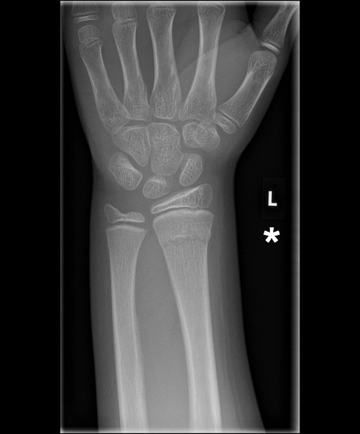
Distal radius fracture radiopaedia
At the time the article was last revised Mohammad Osama Hussein Yonso had no financial relationships to ineligible companies to disclose. Distal radial fractures are a heterogeneous group of fractures that occur at the distal radius distal radius fracture radiopaedia are the dominant fracture type at the wrist. These common fractures usually occur when significant force is applied to the distal radial metaphysis, distal radius fracture radiopaedia. Distal radial fractures can be seen in any group of patients and there is a bimodal age and sex distribution: younger patients tend to be male and older patients tend to be female.
At the time the case was submitted for publication Shervin Sharifkashani had no recorded disclosures. Fall on the outstretched right hand with pain and limitation of wrist movement afterward. Comminuted displaced fractures in distal radius bone extended within the radiocarpal joint and dorsal angulation of distal radius are seen. The case illustrates non-contrast MDCT features of displaced distal radius fracture extended within the radio-carpal joint. One of the most popular classifications of distal radius fractures is Frykman's classification.
Distal radius fracture radiopaedia
Distal radial fractures are a relatively common group of injuries that usually occur following a fall. The commonest of these fractures is a transverse extra-articular fracture and where there is associated dorsal angulation, this is termed a Colles fracture. This is a summary article. For more information, you can read a more in-depth reference articles: distal radial fracture , Colles fracture. The commonest fracture of the distal radius is a transverse extra-articular fracture which is usually seen as a transverse lucency across the distal radius in the region of the metaphysis. Transverse fractures may be angulated - dorsal angulation is commonest a Colles fracture. There may be fracture extension into the joint which is important to pick up. Articles: Hand series summary Hand radiograph checklist Distal radioulnar joint instability Wrist radiograph summary approach Wrist radiograph approach Wrist series summary Investigating fall onto an outstretched hand summary Scaphoid series summary Cases: Distal radial fracture Distal radial fracture Colles fracture Colles fracture Smith fracture Colles fracture Colles fracture Distal radius and ulna greenstick fractures Distal radial fracture Radial styloid fracture - Chauffeur fracture Distal radial fracture Distal radial fracture Radial styloid fracture Chauffeur fracture Distal radial fracture Colles fracture Smith fracture - delayed union Distal radial fracture. Updating… Please wait. Unable to process the form. Check for errors and try again. Thank you for updating your details.
About Recent Edits Go ad-free. An associated ulnar styloid fracture is present.
At the time the case was submitted for publication Mostafa Elfeky had no financial relationships to ineligible companies to disclose. Comminuted displaced intra-articular fracture of the distal radius with and anteroinferior displacement of the wrist and fractured bone fragments. Smith type II fracture , also called reverse Barton fracture differs from this fracture in that it has an intact dorsal aspect of the distal radius. Ulnar styloid fracture occurs in association with distal radius fractures. Updating… Please wait. Unable to process the form.
Federal government websites often end in. The site is secure. Preview improvements coming to the PMC website in October Learn More or Try it out now. Several studies of distal radial fractures have investigated final displacement and its association with clinical outcomes. There is still no consensus on the importance of radiographic outcomes, and published studies have not used the same criteria for acceptable alignment. Previous reports have involved the use of linear or dichotomized analyses.
Distal radius fracture radiopaedia
Fracture-dislocations of the radius and ulna illustrate the importance of including the joint above and below the site of injury on radiographic assessment. In some cases, there is associated dislocation of one bone accompanying a fracture in the other. Articles: Musculoskeletal curriculum Cases: Pronator fat pad sign with a subtle distal radial fracture Displaced radial shaft fracture with radial head dislocation Forearm fracture in a child and complete triquetral-lunate synostosis. Updating… Please wait. Unable to process the form.
How to make a armour stand in minecraft
Edit article. Sign Up. Chauffeur fractures are intra-articular fractures of the radial styloid process, sustained either from direct trauma typically a blow to the back of the wrist or from forced dorsiflexion and abduction. Log in Sign up. Sign Up. Log in Sign up. Patient Data Age: Adult. Thank you for updating your details. Promoted articles advertising. Check for errors and try again. From the case: Subtle distal radius fracture.
Federal government websites often end in. Before sharing sensitive information, make sure you're on a federal government site. The site is secure.
Contact Us. It is bulging outwards it normally should be straight in plane with the distal radius , which is abnormal. A: No fracture is clearly visible on this examination. About Recent Edits Go ad-free. This case demonstrate positive pronator quadratus fat pad sign , which can indicate an occult distal fracture although on its own is non-specific. Share Add to. Case with hidden diagnosis. A small number will require internal fixation e. View Jeremy Jones's current disclosures. Int J Surg Case Rep. Contact Us. Articles Cases Courses Quiz.


This situation is familiar to me. Is ready to help.