Diffusion tensor imaging
At the time the article was last revised Rohit Sharma had no financial relationships to ineligible companies to disclose.
Federal government websites often end in. The site is secure. Diffusion tensor magnetic resonance imaging DTI is a relatively new technology that is popular for imaging the white matter of the brain. The goal of this review is to give a basic and broad overview of DTI such that the reader may develop an intuitive understanding of this type of data, and an awareness of its strengths and weaknesses. We have tried to include equations for completeness but they are not necessary for understanding the paper. Wherever possible, pointers will be provided to more in-depth technical articles or books for further reading.
Diffusion tensor imaging
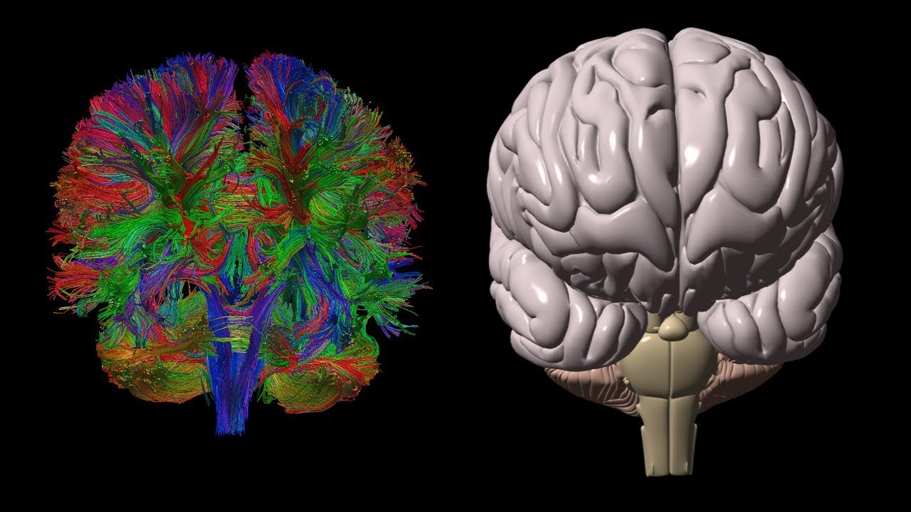
Diffusion-weighted magnetic resonance imaging DWI or DW-MRI is the use of specific MRI sequences as well as software that generates images from the resulting data that uses the diffusion of water molecules to generate contrast in MR images. Molecular diffusion in tissues is not random, but reflects interactions with many obstacles, such as macromolecules , fibers, and membranes. Water molecule diffusion patterns can therefore reveal microscopic details about tissue architecture, either normal or in a diseased state. A special kind of DWI, diffusion tensor imaging DTI , has been used extensively to map white matter tractography in the brain. In diffusion weighted imaging DWI , the intensity of each image element voxel reflects the best estimate of the rate of water diffusion at that location. Because the mobility of water is driven by thermal agitation and highly dependent on its cellular environment, the hypothesis behind DWI is that findings may indicate early pathologic change. A variant of diffusion weighted imaging, diffusion spectrum imaging DSI , [4] was used in deriving the Connectome data sets; DSI is a variant of diffusion-weighted imaging that is sensitive to intra-voxel heterogeneities in diffusion directions caused by crossing fiber tracts and thus allows more accurate mapping of axonal trajectories than other diffusion imaging approaches. Diffusion-weighted images are very useful to diagnose vascular strokes in the brain. It is also used more and more in the staging of non-small-cell lung cancer , where it is a serious candidate to replace positron emission tomography as the 'gold standard' for this type of disease. Diffusion tensor imaging is being developed for studying the diseases of the white matter of the brain as well as for studies of other body tissues see below.
Kochunov, P. Basser PJ, Pajevic S.
Federal government websites often end in. The site is secure. Diffusion tensor imaging DTI is a promising method for characterizing microstructural changes or differences with neuropathology and treatment. The diffusion tensor may be used to characterize the magnitude, the degree of anisotropy, and the orientation of directional diffusion. This review addresses the biological mechanisms, acquisition, and analysis of DTI measurements.
Although DTI and fiber tracking methods are proving valuable in the research realm for groups of subjects, I would caution their application for clinical diagnosis in individual patients. First, considerable variation of regional FA values exists among subjects as a function of age, sex, location, and MR technique. To reduce errors due to crossing, kissing, or branching fibers, a minimum of 20 diffusion directions should be obtained with 64 or more preferred. If FA values are compared to normal data bases, it is critical that multiple comparison e. Bonferroni corrections be performed to avoid spurious conclusions of statistical significance when none exist. Tractography results are even more variable than regional FA measurements and are highly dependent on the exact location of the initial seed points and tracking algorithms employed. An arbitrary starting point must be selected, whose exact location, size and orientation significantly affects results. Tractography algorithms differ in detail between vendors and arbitrary decisions must be made as to when tracking should stop. Termination criteria may be based on FA value terminate tracking when FA falls below a certain value, e.
Diffusion tensor imaging
Diffusion-weighted magnetic resonance imaging DWI or DW-MRI is the use of specific MRI sequences as well as software that generates images from the resulting data that uses the diffusion of water molecules to generate contrast in MR images. Molecular diffusion in tissues is not random, but reflects interactions with many obstacles, such as macromolecules , fibers, and membranes. Water molecule diffusion patterns can therefore reveal microscopic details about tissue architecture, either normal or in a diseased state. A special kind of DWI, diffusion tensor imaging DTI , has been used extensively to map white matter tractography in the brain. In diffusion weighted imaging DWI , the intensity of each image element voxel reflects the best estimate of the rate of water diffusion at that location. Because the mobility of water is driven by thermal agitation and highly dependent on its cellular environment, the hypothesis behind DWI is that findings may indicate early pathologic change.
Bt7274
Cox, R. Each gradient direction applied measures the movement along the direction of that gradient. Please help improve this article by adding citations to reliable sources in this section. Chen, B. Hattingen, E. Ciccarelli, O. Mapping complex tissue architecture with diffusion spectrum magnetic resonance imaging. Brain Stimul. The collection of molecular displacements of this physical property can be described with nine components—each one associated with a pair of axes xx , yy , zz , xy , yx , xz , zx , yz , zy. Unsourced material may be challenged and removed. Wikimedia Commons has media related to Diffusion magnetic resonance imaging.
The precise characterization of cerebral thrombi prior to an interventional procedure can ease the procedure and increase its success.
IEEE Trans. The histogram of each diffusion parameter presents the mean, the peak height and location, values that can be used to compare groups through statistical tests Figure 1M. Tumors are in many instances highly cellular, giving restricted diffusion of water, and therefore appear with a relatively high signal intensity in DWI. Six diffusion-weighted images the minimum number required for tensor calculation. The main issues at this stage are related with the location of the seeding points among different subjects and the fiber tracking tool used, causing variability in the results Burgel et al. However, to date there is no perfect method, and it is unlikely that perfect tractography is possible. Mapping radiation dose distribution on the fractional anisotropy map: applications in the assessment of treatment-induced white matter injury. Several computational methods can be used to perform basic streamline tractography. The first properties they were applied to were those that can be described by a single number, such as temperature. Beaulieu, C. Cases and figures. Lucas, B.


0 thoughts on “Diffusion tensor imaging”